 Explore
 Validate
 Learn
Learn Other assay
Antibody data
- Antibody Data
- Antigen structure
- References [4]
- Comments [0]
- Validations
- Other assay [4]
Submit
Validation data
Reference
Comment
Report error
- Product number
- CSI 001-71-02 - Provider product page

- Provider
- Invitrogen Antibodies
- Product name
- Perlecan Monoclonal Antibody (A71)
- Antibody type
- Monoclonal
- Antigen
- Other
- Description
- CSI 001-71-02 is highly specific for perlecan. There is no evidence for cross-reactivity with other connective tissue proteins (vitronectin, fibronectin, elastin, collagen, laminin). CSI 001-71-02 cross-reacts with human thrombospondin. Other species have not been tested.
- Antibody clone number
- A71
- Concentration
- 1.03 mg/mL
Submitted references Cleavage of the Perlecan-Semaphorin 3A-Plexin A1-Neuropilin-1 (PSPN) Complex by Matrix Metalloproteinase 7/Matrilysin Triggers Prostate Cancer Cell Dyscohesion and Migration.
Differential expression of epithelial basement membrane components nidogens and perlecan in corneal stromal cells in vitro.
Perlecan domain 1 recombinant proteoglycan augments BMP-2 activity and osteogenesis.
Similarity of recombinant human perlecan domain 1 by alternative expression systems bioactive heterogenous recombinant human perlecan D1.
Tellman TV, Cruz LA, Grindel BJ, Farach-Carson MC
International journal of molecular sciences 2021 Mar 22;22(6)
International journal of molecular sciences 2021 Mar 22;22(6)
Differential expression of epithelial basement membrane components nidogens and perlecan in corneal stromal cells in vitro.
Santhanam A, Torricelli AA, Wu J, Marino GK, Wilson SE
Molecular vision 2015;21:1318-27
Molecular vision 2015;21:1318-27
Perlecan domain 1 recombinant proteoglycan augments BMP-2 activity and osteogenesis.
Decarlo AA, Belousova M, Ellis AL, Petersen D, Grenett H, Hardigan P, O'Grady R, Lord M, Whitelock JM
BMC biotechnology 2012 Sep 11;12:60
BMC biotechnology 2012 Sep 11;12:60
Similarity of recombinant human perlecan domain 1 by alternative expression systems bioactive heterogenous recombinant human perlecan D1.
Ellis AL, Pan W, Yang G, Jones K, Chuang C, Whitelock JM, DeCarlo AA
BMC biotechnology 2010 Sep 9;10:66
BMC biotechnology 2010 Sep 9;10:66
No comments: Submit comment
Supportive validation
- Submitted by
- Invitrogen Antibodies (provider)
- Main image

- Experimental details
- Figure 2 ELISA identifies only Pln.D1 but not Pln.D3-5 in enriched rhPln.198 . The enriched high M r rhPln.198 synthesized by HEK 293 cells was used to coat microtiter wells that were subsequently blocked then incubated with 1.7 mug/ml of different primary antibodies as depicted in the legend. ELISA was completed as described in Methods. Background signal for each antibody binding to uncoated wells was subtracted to produce the net signal. Inset: ELISA data showing negligible mAb A76 reactivity against rhPln.247 and strong reactivity with full-length endothelial perlecan (purified as previously described [ 53 ]), while mAb A71 reacted strongly to both the native perlecan and the truncated recombinant. Net absorbance minus buffer-coated wells represented.

- Submitted by
- Invitrogen Antibodies (provider)
- Main image

- Experimental details
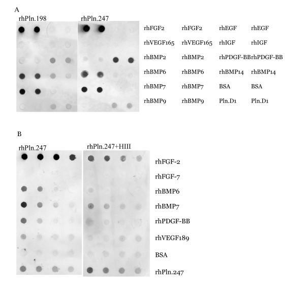
- Figure 8 Immunoblot analysis of growth factor binding to Pln.198 and Pln.247 . 500 ng growth factors were bound to nitrocellulose, the blots were blocked, and then incubated in either 1 ug/ml Pln.198 or Pln.247 (panel A) or rhPln.247 pre-digested with heparinase III (panel B). Detection was performed with anti-perlecan domain 1 primary antibody CSI 001-71. Note that while both the background BSA signal and the internal control rhPln.247 signals are slightly higher in panel B (bottom two samples), a decrease in the growth factor binding to Pln.247 was present after digestion.
- Submitted by
- Invitrogen Antibodies (provider)
- Main image
- Experimental details
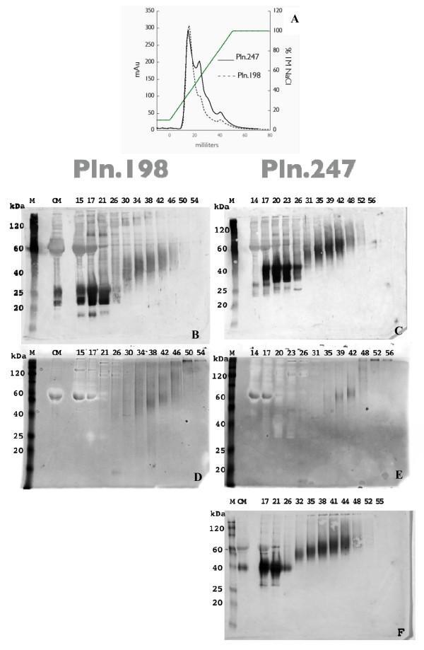
- Figure 1 Anion Exchange Chromatography of rhPln.247 conditioned media (CM) . A: Chromatogram of elution profile for rhPln.198-Ad and rhPln.247-Ad CM expressed from HEK 293 cells; B and C: Western blots of anion exchange fractions with mAb CSI 001-71 recognizing the Pln.D1 core; D and E: Stains-All analysis of HEK 293 fractions; F: Fractions from HUVEC synthesis. Panels B and D represent rhPln.198 fractionation; panels C, E, F represent rhPln.247 fractionation. (M) Markers; (CM) Conditioned medium undiluted from cells. Similar data were generated using plasmid expression of pln.247 (not shown).
- Submitted by
- Invitrogen Antibodies (provider)
- Main image

- Experimental details
- Figure 4 Perlecan immunocytochemistry of corneal stromal cells cultured under different conditions. Perlecan (red) was most highly expressed in localized intracellular structures in keratocytes whereas in the myofibroblasts perlecan was detected more diffusely throughout the cell (white arrowheads). Fibroblasts had little, if any, detectable expression of perlecan protein. The isotypic control immunoglobulin (IgG) did not yield staining under any of the culture conditions, as is shown for myofibroblasts. Magnification=400X.
